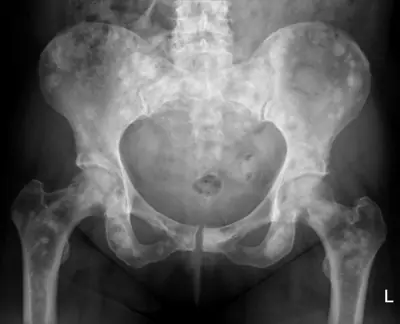
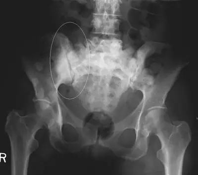
Case: Bone & joint disease radiograph #119640855
Bone & joint disease Intermediate
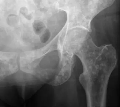
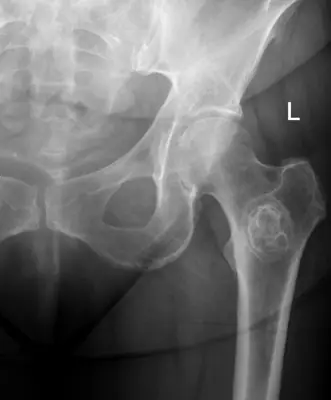
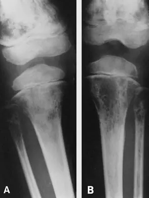
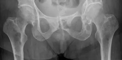
Read this radiograph systematically before revealing the answer. What is the main abnormality?
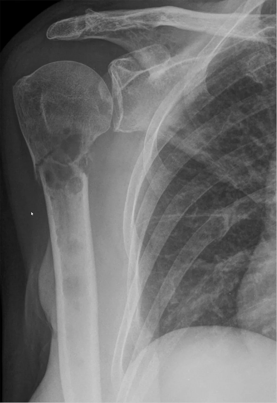
Image: Hellerhoff via Wikimedia Commons · CC BY-SA 4.0 · source · resized and converted to WebP
Tip: scroll or pinch to zoom, drag to pan, and try Invert.
Answer: Bone metastases
What to look for in bone metastases
- Lytic (destructive), sclerotic (dense) or mixed lesions, often multiple.
- Typical sites are red-marrow areas: spine, pelvis, ribs, skull and proximal long bones.
- A missing pedicle on the spine AP view ('winking owl') is a classic sign; check for pathological fracture.
Description by the image author (may be in another language): Pathological fracture of right proximal humerus in a patient with metastatic breast cancer. Other osteolytic lesions are also seen at glenoid cavity and several ribs. Pathological fractures are also seen at the ribs.
Educational case. The diagnosis comes from the image's documented source; the "what to look for" list describes the condition in general and is not a report of this individual image.