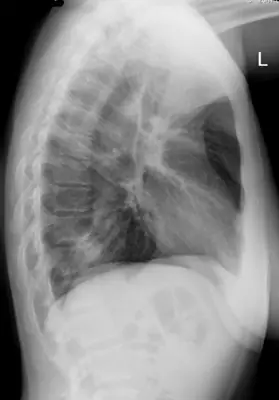
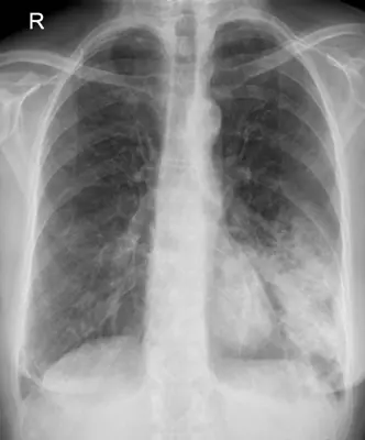
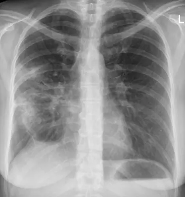
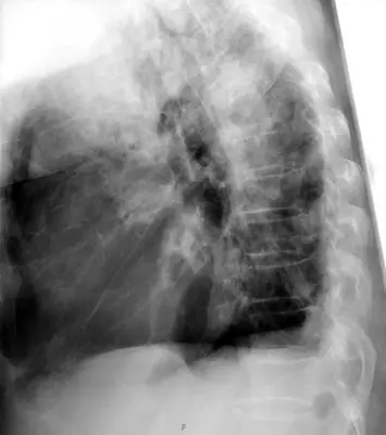
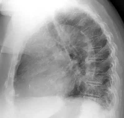
Case: Chest radiograph #76593239
Chest Beginner
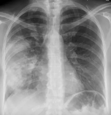
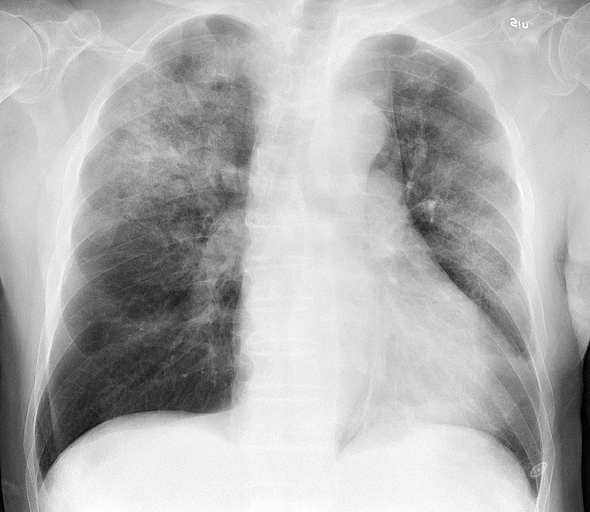
Read this radiograph systematically before revealing the answer. What is the main abnormality?
Tip: scroll or pinch to zoom, drag to pan, and try Invert.
Answer: Pneumonia (consolidation)
What to look for in pneumonia (consolidation)
- Dense RUL opacity bounded by the horizontal fissure.
- Air bronchograms.
- No volume loss.
Description by the image author (may be in another language): Chest radiograph of an 88 year old man, about one week after onset of fever, fatigue and mild coughing. Lab tests detected both Influenza A virus and Haemophilus influenzae . It shows multifocal, patchy consolidation, mainly in the right upper lobe.
Educational case. The diagnosis comes from the image's documented source; the "what to look for" list describes the condition in general and is not a report of this individual image.