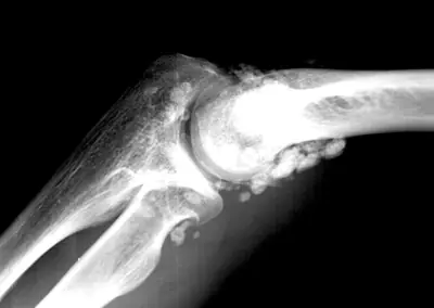
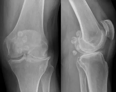
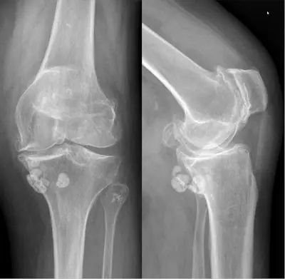
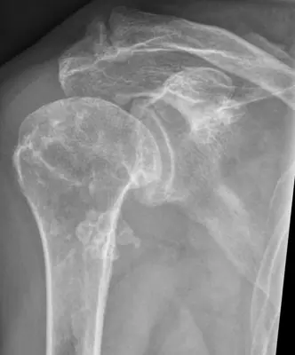
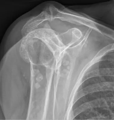
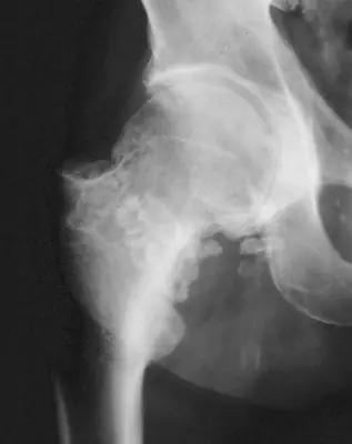
Case: Bone & joint disease radiograph #118606331
Bone & joint disease Advanced
Read this radiograph systematically before revealing the answer. What is the main abnormality?
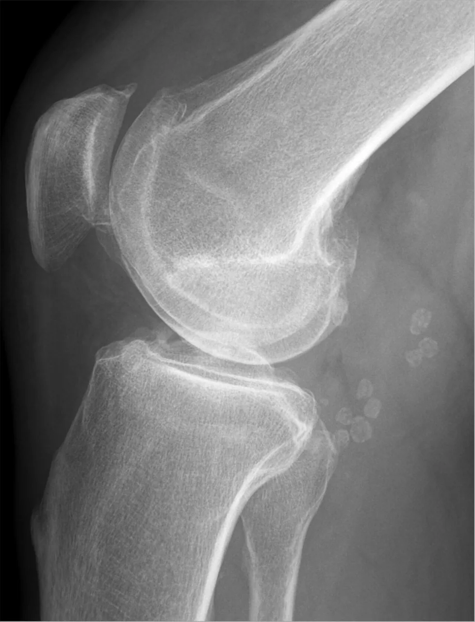
Image: Hellerhoff via Wikimedia Commons · CC BY-SA 4.0 · source · resized and converted to WebP
Tip: scroll or pinch to zoom, drag to pan, and try Invert.
Answer: Synovial chondromatosis
What to look for in synovial chondromatosis
- Multiple small, similar-sized calcified or ossified loose bodies within a joint.
- Knee and hip are the commonest sites.
- Joint space is often preserved early, which helps distinguish it from osteoarthritis with loose bodies.
Description by the image author (may be in another language): Synovial chondromatosis (a collection of loose cartilagenous bodies which may or may not calcified) occuring within Baker's cyst (fluid-filled lesion lined by synovium, located within the popliteal fossa, that communicates with the knee joint).
Educational case. The diagnosis comes from the image's documented source; the "what to look for" list describes the condition in general and is not a report of this individual image.