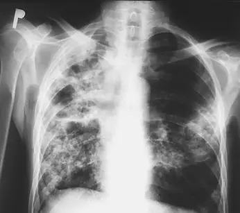
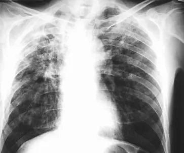
Case: Chest radiograph #880747
Chest Intermediate
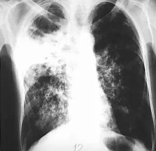
Read this radiograph systematically before revealing the answer. What is the main abnormality?
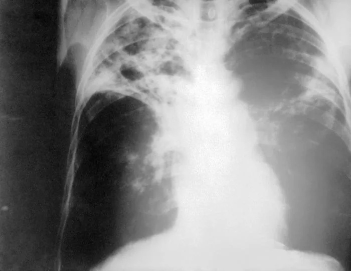
Image: Photo Credit: Content Providers(s): CDC via Wikimedia Commons · Public domain · source · resized and converted to WebP
Tip: scroll or pinch to zoom, drag to pan, and try Invert.
Answer: Pulmonary tuberculosis
What to look for in pulmonary tuberculosis
- Upper-zone consolidation with cavitation and volume loss.
Description by the image author (may be in another language): An anteroposterior X-ray of a patient diagnosed with advanced bilateral pulmonary tuberculosis. This AP X-ray of the chest reveals the presence of bilateral pulmonary infiltrate, and „caving formation“ present in the right apical region. The diagnosis is far-advanced tuberculosis.
Educational case. The diagnosis comes from the image's documented source; the "what to look for" list describes the condition in general and is not a report of this individual image.