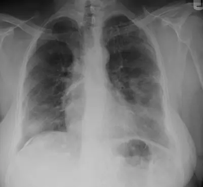
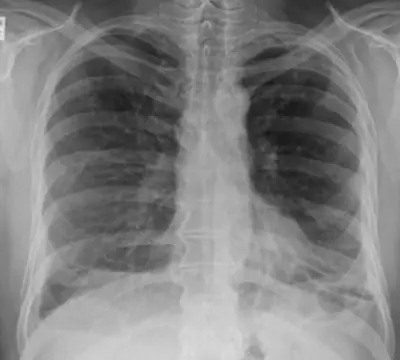
Case: Chest radiograph #729018
Chest Intermediate
Read this radiograph systematically before revealing the answer. What is the main abnormality?
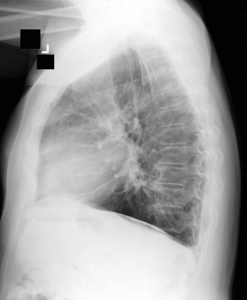
Image: User Clinical Cases on en.wikipedia via Wikimedia Commons · CC BY-SA 2.5 · source · resized and converted to WebP
Tip: scroll or pinch to zoom, drag to pan, and try Invert.
Answer: Asbestos-related pleural disease
What to look for in asbestos-related pleural disease
- Calcified holly-leaf plaques.
- Plaques on the diaphragm.
Description by the image author (may be in another language): Chest X-ray in asbestosis shows plaques above diaphragm Source: Early Asbestosis in a Retired Pipe Fitter http://clinicalcases.blogspot.com/2004/03/early-asbestosis-in-retired-pipe.html Clinical_Cases : I made the photo myself, licensed under Creative Commons license.
Educational case. The diagnosis comes from the image's documented source; the "what to look for" list describes the condition in general and is not a report of this individual image.