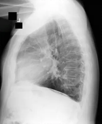
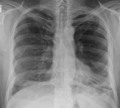
Case: Chest radiograph #82807623
Chest Intermediate
Read this radiograph systematically before revealing the answer. What is the main abnormality?
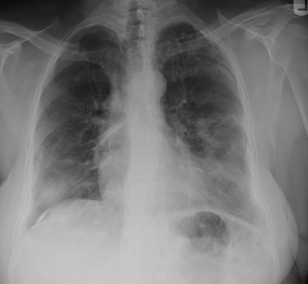
Image: Yale Rosen from USA via Wikimedia Commons · CC BY-SA 2.0 · source · resized and converted to WebP
Tip: scroll or pinch to zoom, drag to pan, and try Invert.
Answer: Asbestos-related pleural disease
What to look for in asbestos-related pleural disease
- Calcified holly-leaf plaques.
- Plaques on the diaphragm.
Description by the image author (may be in another language): There is a calcified pleural plaque on the lateral surface of the right hemodiaphragm (See Note). A needle core biopsy of the nodular infiltrate in the right lower lobe showed cryptococcosis and asbestos bodies. There is a history of significant asbestos exposure.
Educational case. The diagnosis comes from the image's documented source; the "what to look for" list describes the condition in general and is not a report of this individual image.