Pelvic phleboliths on X‑ray: 5 real cases
Real, openly licensed radiographs showing pelvic phleboliths. Study what to look for, then open each case and read it yourself before revealing the answer.
What to look for
- Small, round, smooth calcifications low in the pelvis, often with a lucent centre.
- They are calcified venous thrombi and are very common incidental findings.
- The main differential is a distal ureteric stone, which tends to be more irregular and lies along the ureteric course.
Learn: Calcification and stones
Quiz yourself on abdomen images
Cases
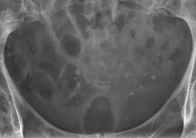 Case #101675200Abdomen · Intermediate
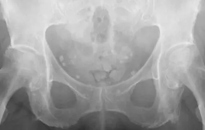 Case #124127877Abdomen · Intermediate
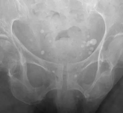 Case #125453404Abdomen · Intermediate
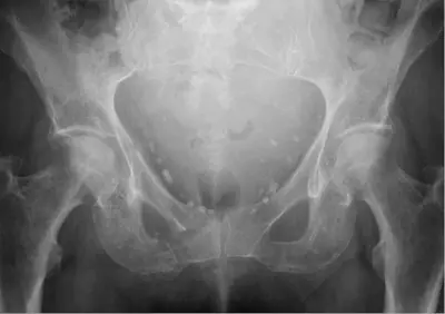 Case #110522503Abdomen · Intermediate
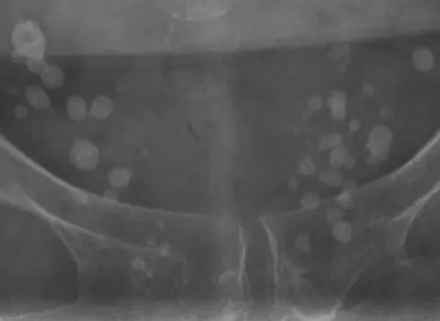 Case #122454053Abdomen · Intermediate
Other abdomen diagnoses
- Bowel obstruction
- Calcified uterine fibroid
- Chilaiditi sign (colon interposed under the diaphragm)
- Foreign body (ingested or inserted)
- Intrauterine contraceptive device (IUCD)
- Pneumoperitoneum (free intraperitoneal gas)
- Urinary tract calculi (kidney or ureteric stones)
- Volvulus
Diagnoses come from each image's documented source. Credits and licences are shown on each case and on the image credits page.