Stress fracture on X‑ray: 3 real cases
Real, openly licensed radiographs showing stress fracture. Study what to look for, then open each case and read it yourself before revealing the answer.
What to look for
- Often invisible at first; later a faint lucent line, band of sclerosis or periosteal new bone.
- Typical sites: metatarsal shafts, tibia, calcaneus and femoral neck.
- MRI is more sensitive early; a normal film does not exclude it.
Quiz yourself on lower limb images
Cases
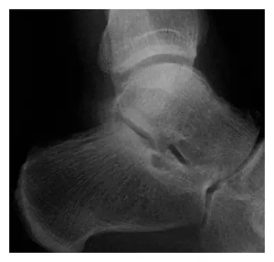 Case #67079391Lower limb · Advanced
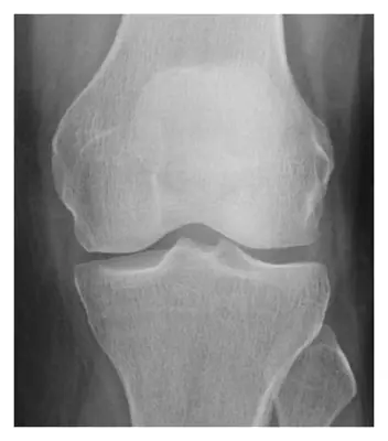 Case #67086624Lower limb · Advanced
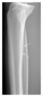 Case #67086626Lower limb · Advanced
Other lower limb diagnoses
- Acetabular fracture
- Ankle fracture (lateral malleolus)
- Bimalleolar ankle fracture
- Calcaneal fracture
- Calcaneal spur
- Femoral shaft fracture
- Fifth metatarsal fracture (Jones / base)
- Hip (proximal femoral) fracture
- Hip dislocation
- Knee joint effusion
- Patellar dislocation
- Patellar fracture
- Pellegrini–Stieda lesion
- Tibial (and fibular) shaft fracture
- Tibial plateau fracture
Diagnoses come from each image's documented source. Credits and licences are shown on each case and on the image credits page.