Thoracic aortic aneurysm on X‑ray: 5 real cases
Real, openly licensed radiographs showing thoracic aortic aneurysm. Study what to look for, then open each case and read it yourself before revealing the answer.
What to look for
- Widened or abnormally prominent aortic contour (knuckle or descending aorta).
- Mediastinal widening that follows the course of the aorta.
- Curvilinear wall calcification may outline the dilated aorta.
- Plain film cannot exclude or characterise aneurysm or dissection; CT angiography is required.
Cases
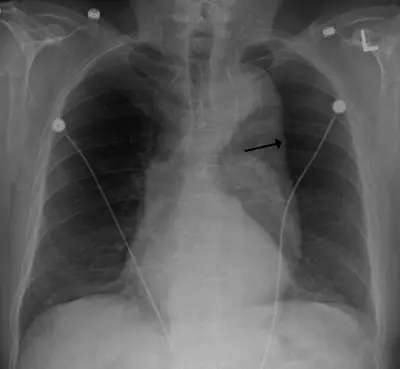 Case #12186251Chest · Advanced
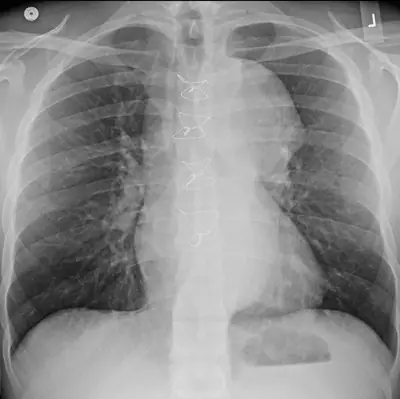 Case #49082190Chest · Advanced
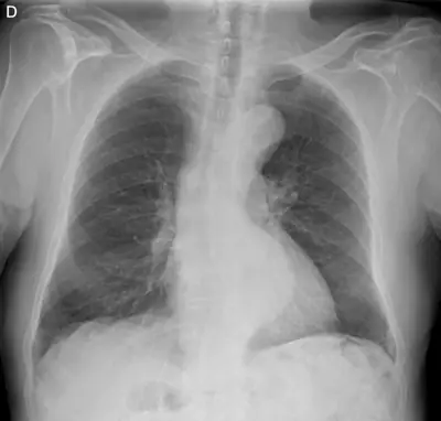 Case #162791180Chest · Advanced
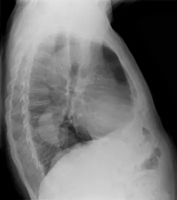 Case #162791182Chest · Advanced
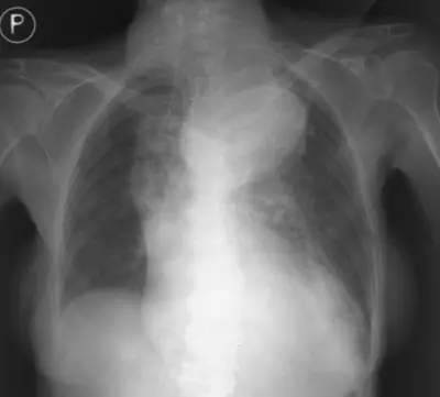 Case #47020165Chest · Advanced
Other chest diagnoses
- Asbestos-related pleural disease
- COVID-19 pneumonia
- Cardiomegaly
- Cervical rib
- Diaphragmatic hernia
- Emphysema / COPD (hyperinflation)
- Hiatus hernia
- Lung abscess
- Lung cancer (mass)
- Lung collapse (atelectasis)
- Normal chest radiograph
- Pectus excavatum
- Pericardial effusion
- Pleural effusion
- Pneumocystis pneumonia (PCP)
- Pneumomediastinum
- Pneumonia (consolidation)
- Pneumothorax
- Pulmonary oedema (heart failure)
- Pulmonary tuberculosis
- Sarcoidosis
- Silicosis / pneumoconiosis
Diagnoses come from each image's documented source. Credits and licences are shown on each case and on the image credits page.