Pectus excavatum on X‑ray: 3 real cases
Real, openly licensed radiographs showing pectus excavatum. Study what to look for, then open each case and read it yourself before revealing the answer.
What to look for
- Indistinct right heart border that mimics right middle lobe disease.
- Heart displaced to the left and sometimes rotated.
- Anterior ribs sloping steeply downwards (horizontal posterior, vertical anterior ribs).
- On the lateral film, the sternum is depressed posteriorly towards the spine.
Cases
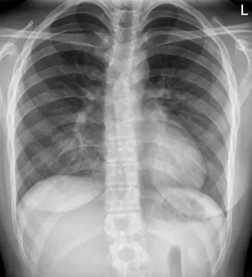 Case #12429301Chest · Intermediate
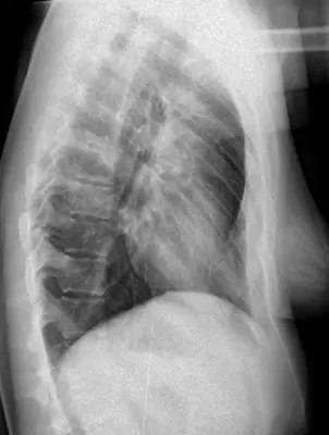 Case #12429320Chest · Intermediate
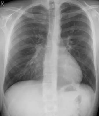 Case #20314526Chest · Intermediate
Other chest diagnoses
- Asbestos-related pleural disease
- COVID-19 pneumonia
- Cardiomegaly
- Cervical rib
- Diaphragmatic hernia
- Emphysema / COPD (hyperinflation)
- Hiatus hernia
- Lung abscess
- Lung cancer (mass)
- Lung collapse (atelectasis)
- Normal chest radiograph
- Pericardial effusion
- Pleural effusion
- Pneumocystis pneumonia (PCP)
- Pneumomediastinum
- Pneumonia (consolidation)
- Pneumothorax
- Pulmonary oedema (heart failure)
- Pulmonary tuberculosis
- Sarcoidosis
- Silicosis / pneumoconiosis
- Thoracic aortic aneurysm
Diagnoses come from each image's documented source. Credits and licences are shown on each case and on the image credits page.