Hallux rigidus (first MTP osteoarthritis) on X‑ray: 5 real cases
Real, openly licensed radiographs showing hallux rigidus (first mtp osteoarthritis). Study what to look for, then open each case and read it yourself before revealing the answer.
What to look for
- Joint space narrowing of the first metatarsophalangeal joint.
- Dorsal osteophytes on the first metatarsal head, best seen on the lateral view.
- Subchondral sclerosis and cysts; alignment is often preserved (unlike hallux valgus).
Quiz yourself on bone & joint disease images
Cases
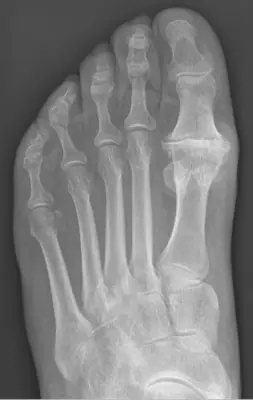 Case #48407447Bone & joint disease · Intermediate
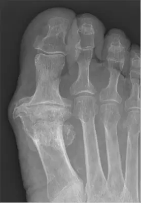 Case #105448936Bone & joint disease · Intermediate
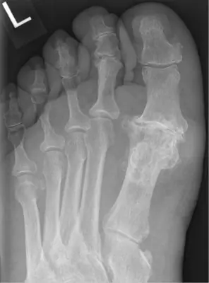 Case #105610628Bone & joint disease · Intermediate
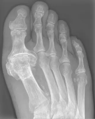 Case #132319338Bone & joint disease · Intermediate
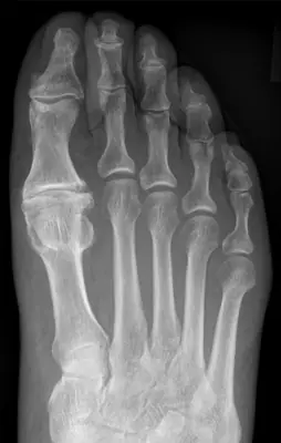 Case #145835713Bone & joint disease · Intermediate
Other bone & joint disease diagnoses
- Avascular necrosis of the femoral head
- Bone metastases
- Enchondroma
- Freiberg disease
- Gout
- Hallux valgus (bunion)
- Heterotopic ossification
- Knee osteoarthritis
- Multiple myeloma
- Neuropathic (Charcot) arthropathy
- Non-ossifying fibroma
- Osteoarthritis
- Osteochondroma (exostosis)
- Osteoma
- Osteomyelitis
- Osteoporosis
- Rheumatoid arthritis
- Synovial chondromatosis
Diagnoses come from each image's documented source. Credits and licences are shown on each case and on the image credits page.