Osteoma on X‑ray: 10 real cases
Real, openly licensed radiographs showing osteoma. Study what to look for, then open each case and read it yourself before revealing the answer.
What to look for
- A well-defined, very dense (ivory) bony lesion.
- Most often arises from the skull vault or the paranasal sinuses (especially frontal).
- Benign; multiple osteomas raise the possibility of Gardner syndrome.
Quiz yourself on bone & joint disease images
Cases
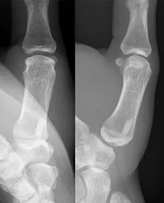 Case #169481829Bone & joint disease · Intermediate
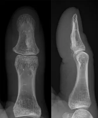 Case #156247345Bone & joint disease · Intermediate
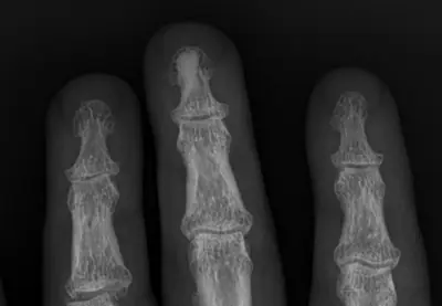 Case #134044372Bone & joint disease · Intermediate
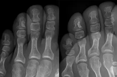 Case #169174013Bone & joint disease · Intermediate
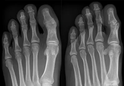 Case #165599280Bone & joint disease · Intermediate
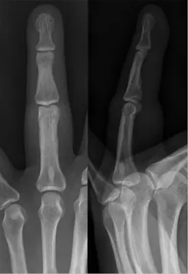 Case #99897726Bone & joint disease · Intermediate
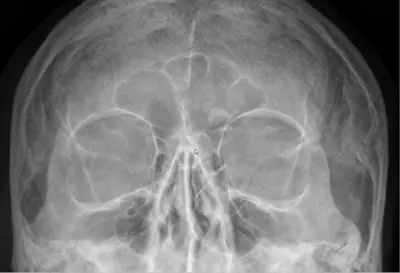 Case #97704527Bone & joint disease · Intermediate
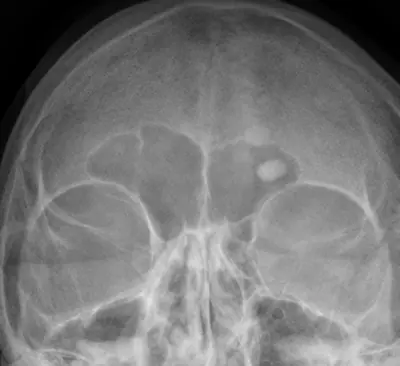 Case #13735210Bone & joint disease · Intermediate
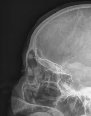 Case #13735291Bone & joint disease · Intermediate
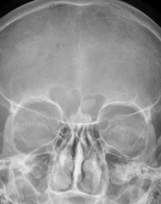 Case #18224362Bone & joint disease · Intermediate
Other bone & joint disease diagnoses
- Avascular necrosis of the femoral head
- Bone metastases
- Enchondroma
- Freiberg disease
- Gout
- Hallux rigidus (first MTP osteoarthritis)
- Hallux valgus (bunion)
- Heterotopic ossification
- Knee osteoarthritis
- Multiple myeloma
- Neuropathic (Charcot) arthropathy
- Non-ossifying fibroma
- Osteoarthritis
- Osteochondroma (exostosis)
- Osteomyelitis
- Osteoporosis
- Rheumatoid arthritis
- Synovial chondromatosis
Diagnoses come from each image's documented source. Credits and licences are shown on each case and on the image credits page.